

Scientists at Harvard University have developed a multicolor microscopy technique that bridges a long-lasting micro-imaging gap.
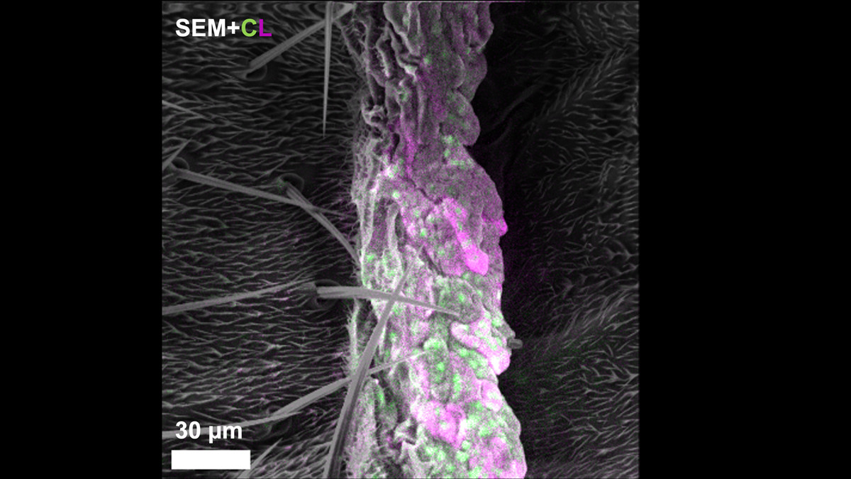
Multicolor EM image of a fungus-infected fly. The nuclei (green) and mitochondria (purple) of the fungus are visible along with the surface structures (SEM) of the fungus-infected fly. /Biophysical Society
This technique allows simultaneous observation of cellular structures and specific protein locations at nanometer-scale resolution.
The research was presented at the 70th Biophysical Society Annual Meeting held in San Francisco from last Saturday.
The technology addresses a longstanding limitation in biological imaging, where scientists previously had to choose between visualizing detailed cell structures or identifying specific molecules.
By enabling both capabilities within a single imaging process, the method provides new opportunities to study biological processes ranging from cellular signaling to molecular cluster organization.
The Harvard team instead employed a single electron beam to obtain structural and molecular information simultaneously, eliminating the need for dual imaging workflows.
The researchers developed a specialized probe that attaches to target proteins and emits visible light through cathodoluminescence when stimulated by the electron beam.
The technique has been validated in mammalian cells and biological tissues, including fungus-infected fruit flies.
Currently limited to two-dimensional imaging, the research team plans to extend the technology to three-dimensional cellular reconstruction using cryo-electron microscopy.